X-ray methods of research

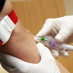
X-ray methods of examination help to diagnose many diseases of internal organs and systems . A nurse, depending on the position and field of work, one way or another is faced with x-ray diagnostics.
The tasks of the nurse may be to ensure the preparation of the patient for x-ray examinations, help the doctor during the procedure, or directly conduct it (X-ray laboratory assistant).
The study of organs using X-rays is based on their different permeability to tissues of different densities. X-rays, when projected onto X-ray film after passing through the internal organs of a person, give a negative image of the organs under study.
There are different types of X-ray studies, which we will consider.
X-ray - obtaining a negative image of the organ under study on photographic film. This method is based on obtaining x-ray images in various projections and assessing the state of the organ from the obtained images.
Tomography— layer-by-layer radiography of the studied organ. Studies are carried out on a classic or spiral computed tomography.
Fluoroscopy - examination of the organ under study behind a special x-ray screen. The method allows to study the anatomical features of the organ and evaluate its functional state.
Fluorography is a method of obtaining reduced-sized images of the chest organs. At the same time, the number of X-rays passing through the examined patient is also reduced.
Bone tissue is examined absolutely reliably, because has a dense structure. Some internal organs, consisting of dense parenchymal tissue, are also well defined on x-rays. To study hollow organs, special techniques are used to make them more accessible for research - the use of radiopaque substances (drugs).
Modern X-ray equipment makes it possible to obtain a spatial image of an organ, a video recording of its work, and also to highlight the part of the organ that is of interest to the doctor.
Some X-ray methods of research for various organs by systems
Respiratory system:
- plain radiography and fluoroscopy of the chest organs without preliminary contrasting;
- contrast bronchography and bronchoscopy.
The cardiovascular system:
- radiography of the heart in several projections;
- teleradiography of the heart;
- phlebography;
- plain radiography and fluoroscopy of the chest.
Digestive system:
- survey radiography and fluoroscopy of the abdominal organs without preliminary contrasting;
- radiography and fluoroscopy of the stomach and duodenum using a contrast agent;
- irrigoscopy and irrigography - examination of the large intestine;
- cholecystography and cholecystoscopy using a contrast agent - examination of the gallbladder;
- contrast cholegraphy - a study of the bile ducts and gallbladder using the contrast method.
Urinary system:
- survey radiography and fluoroscopy of the entire system without preliminary contrasting;
- intravenous urography - examination of the kidneys, ureters, bladder with preliminary contrasting.
For a nurse to work as an X-ray laboratory assistant, it is necessary to obtain specialization by completing a training course with subsequent testing.
The ward nurse needs to clearly know the algorithm for preparing for all methods of X-ray examinations.